Sanakirja
Tekoälykääntäjä

Kuvat
15

| Kieli | Käännökset |
|---|---|
| espanja | retina |
| esperanto | retino |
| hollanti | netvlies, retina |
| italia | retina |
| japani | 網膜 (mōmaku) |
| kreikka | αμφιβληστροειδής (amfivlistroeidís) |
| latvia | tīklene |
| portugali | retina |
| puola | siatkówka |
| ranska | rétine |
| ruotsi | näthinna, retina |
| saksa | Netzhaut, Retina |
| suomi | verkkokalvo, retina |
| turkki | ağ kat, ağ tabaka |
| tšekki | sítnice |
| unkari | recehártya, retina |
| venäjä | сетчатка (settšatka), ретина (retina) |
Määritelmät
Substantiivi
- (ophthalmology) The thin layer of cells at the back of the eyeball that contains rods and cones sensitive to light, which trigger nerve impulses that pass via the optic nerve to the brain, where a visual image is formed.
Taivutusmuodot
| Monikko | retinas |
| Monikko | retinae |
| Monikko | retinæ (vanhentunut) |
Luokat
(ophthalmology) The thin layer of cells at the back of the eyeball that contains rods and cones sensitive to light, which trigger nerve impulses that pass via the optic nerve to the brain, where a visual image is formed.
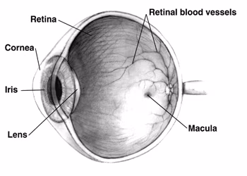
Illustration showing parts of the human eye, including the retina.
(ophthalmology) The thin layer of cells at the back of the eyeball that contains rods and cones sensitive to light, which trigger nerve impulses that pass via the optic nerve to the brain, where a visual image is formed.

an orthographic cross-section of the layers of the human retina labeling various elements. Light comes from top right
(ophthalmology) The thin layer of cells at the back of the eyeball that contains rods and cones sensitive to light, which trigger nerve impulses that pass via the optic nerve to the brain, where a visual image is formed.

Rods, cones, and nerve layers in the retina: The front (anterior) of the eye is on the left. Light (from the left) passes through several transparent nerve layers to reach the rods and cones (far right). Chemical changes in the rods and cones send a signal back to the nerves. The signal goes first to the bipolar and horizontal cells (yellow layer), then to the amacrine cells and ganglion cells (purple layer), then to the optic nerve fibres. The signals are processed in these layers. First, the signals start as raw outputs of points in the rod and cone cells. Then, the nerve layers identify simple shapes, such as bright points surrounded by dark points, edges, and movement. (Based on a drawing by Ramón y Cajal, 1911)